

Biomechanical Assessment
This encompasses joint range of motion and quality of motion study, examination of deformity, structure and alignment. The best results are achieved whilst the patient lies supine (face up) or prone (face down) or whilst standing.Gait Assessment
Information is obtained by observing gait variations or abnormalities. With variation alignment and/or function of the shoulders, spine, pelvis, hip, knee, ankle and foot are observed.This may involve the assessment of the pulses of the foot, blood pressure readings (if required), arterial health (i.e. via Doppler if required), skin temperature, trophic changes (i.e. quality of skin and nails), presence or absence of sensory or motor neuropathy and the presence or absence of deformity. This assessment is important for diabetic patients to determine their risk of developing a foot complication such as ulceration and/or infection. Patients are classified as being at low, medium or high risk.
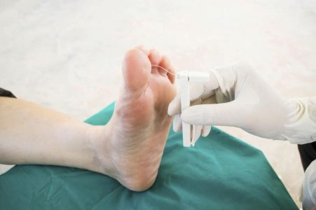
Monofilament Testing
Monofilament testing is a cheap and highly effective way of detecting sensory loss. A loss of foot sensation is an important predictor for ulceration in the diabetic patient.
Plain film radiographs (x-rays), ultrasound, magnetic resonance imaging (MRI), duplex ultrasound (arterial or venous) and ultrasound are common forms of medical imaging. Our practitioners commonly utilise these techniques and other methods of imaging to assess foot, ankle and lower limb pathology.
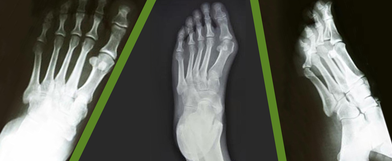
Plain Film Radiographs (x-rays)
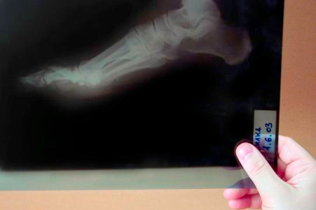
These are by far the most common form of medical imaging used by podiatrists and podiatric surgeons. They are used to assess the bone and joint health of the foot ankle. When necessary measurements can be taken to compare the extent of structural variation or deformity with the normal population. Such measurments can be a valuable aid when determining the best form of treatment, whether non-surgical or surgical.Plain film radiographs (x-rays) are the most common form of imaging for foot and ankle disorders.
Ultrasound (musculoskeletal)
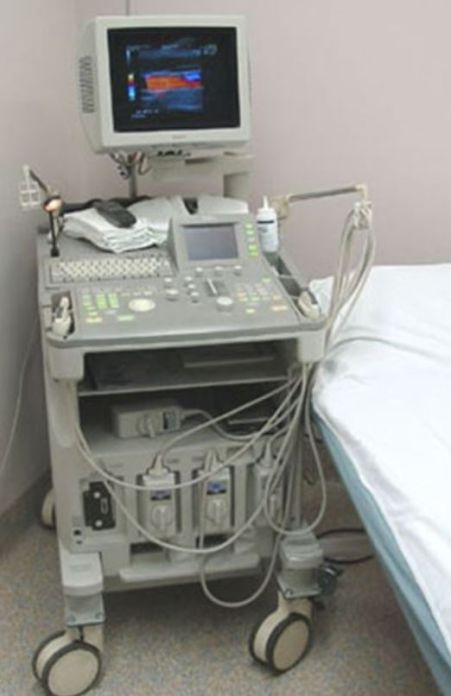
Ultrasound technology is used to assess for soft tissue disorders such as tendon injury, cysts, ganglions, bursitis and neuromas (Ultrasound - Normal Foot and Ultrasound - Pathology Foot). Ultrasound is often combined with injection therapy (a corticosteroid combined with a local anaesthetic) as the operator can see the damaged area whilst it is being injected. Such injections can be very useful for providing short term and at times long term pain relief. Ultrasound is highly operator dependent and as such is highly versatile.
Ultrasound Machine: Musculoskeletal ultrasound is an excellent imaging modality for examining soft tissue disorders (including bumps and lumps) of the foot. Corticosteroid injections used to treat painful conditions of the foot are commonly performed under the guidance of ultrasound
MRI (magnetic resonance imaging)
This is the gold standard of musculoskeletal imaging and is useful in complex foot and ankle conditions, especially when multiple structures are affected, such as soft tissue and bone injury following trauma. MRI is commonly used for a diabetic patient with a suspected soft tissue and/or bone infection. MRI is very useful for chronic pain in which clinical diagnosis has proven difficult.